Difference: GMT_3dus (1 vs. 3)
Revision 3
15 Jul 2014 - gmt11
Revision 2
15 Jul 2014 - gmt11
| Line: 1 to 1 | ||||||||
|---|---|---|---|---|---|---|---|---|
| ||||||||
| Changed: | ||||||||
| < < | Dr Graham Treece, Department of Engineering | |||||||
| > > |
Dr Graham Treece, Department of Engineering | |||||||
|
| ||||||||
| Line: 11 to 11 | ||||||||
Overview | ||||||||
| Changed: | ||||||||
| < < |
 | |||||||
| > > |
 | |||||||
The objectives of this project are:
| ||||||||
| Line: 27 to 28 | ||||||||
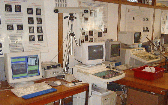
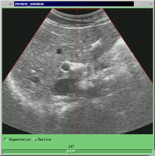
| We have developed a high-resolution version of the Stradx 3D ultrasound acquisition system. Three elements of the existing design were enhanced to improve the accuracy and resolution: the 2D ultrasound images come from a Dynamic Imaging `Diasus' machine with resolution up to 0.05mm; these images are digitally transferred from the ultrasound machine to a computer; and the position sensing is performed using an optical rather than a magnetic system. | ||||||||
| Changed: | ||||||||
| < < |
 | |||||||
| > > |
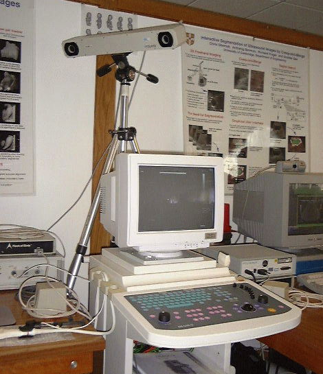 | |||||||
| A high bandwidth ethernet link is used to transfer the digital ultrasound data from the Diasus machine to the computer running Stradx. This enables us to eliminate errors introduced by the PAL image encoding, and the A-D converter in the frame grabber. | ||||||||
| Line: 41 to 44 | ||||||||
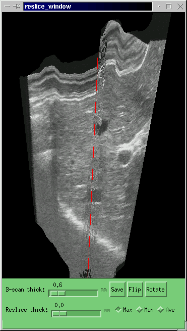
| We therefore correct pressure-induced artefacts using sequential image correlation techniques. A rigid in-plane translation and non-rigid depth compression are applied to each B-scan, so as to maximise the correlation between successive images. Once this has been done to all the B-scans, the 3D reconstruction will be locally smooth but may suffer from large-scale error accumulation. The entire data set is therefore subjected to one final, rigid affine transformation, so that the first and last B-scans in the sequence coincide with their externally measured positions. The image-based registration ensures that the reconstruction is locally smooth, while the position sensor readings guarantee the large-scale positional accuracy of the data. | ||||||||
| Changed: | ||||||||
| < < |
  | |||||||
| > > |
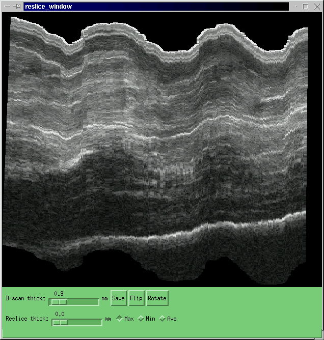 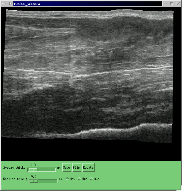 | |||||||
| The figure above shows the original (left) and corrected (right) reslice through a 3DUS volumetric data set of the forearm. | ||||||||
| Changed: | ||||||||
| < < |
  | |||||||
| > > |
 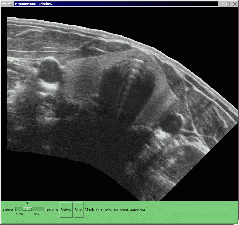 | |||||||
| This procedure is also of benefit for panoramic data sets, for instance this of both lobes of the thyroid. | ||||||||
| Added: | ||||||||
| > > | ||||||||
Convex ProbesWe are also investigating the application of the probe pressure correction algorithm described above to convex probes. For instance, the following figure shows one B-scan from a sequence of scans of the liver. The left hand image is the original: the right hand image has been corrected for convex probe pressure. | ||||||||
| Line: 52 to 56 | ||||||||
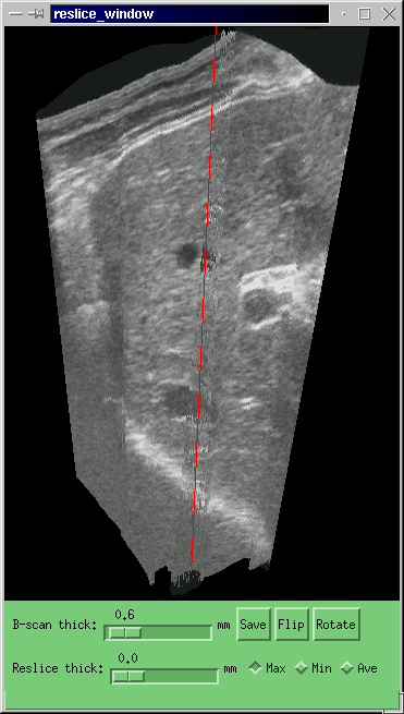
| We are also investigating the application of the probe pressure correction algorithm described above to convex probes. For instance, the following figure shows one B-scan from a sequence of scans of the liver. The left hand image is the original: the right hand image has been corrected for convex probe pressure. | ||||||||
| Changed: | ||||||||
| < < |
  | |||||||
| > > |
  | |||||||
| The effect of this correction on the whole data set can be seen in a reslice through all the original scans. Once again, the corrected image is on the right. | ||||||||
| Changed: | ||||||||
| < < |
  | |||||||
| > > |
  | |||||||
Related Publications | ||||||||
| Line: 71 to 75 | ||||||||
A complete list of the on-line publications from our group is available here.
People / Contacts | ||||||||
| Changed: | ||||||||
| < < |
  | |||||||
| > > |
  | |||||||