
|
Department of Engineering |
Ear surgery, such as cochlear implantation but other surgeries also, involves negotiating many potential hazards. These include not drilling accidently into the facial nerve (which moves the face), the inner ear (causes deafness and balance loss if hit), the sigmoid sinus (copious bleeding) or the dura (causes brain fluid leakage), to name but a few. Structures are packed together tightly and covered in bone, so not visible untill the bone is drilled away. This project aims to help the surgeon stay oriented during the surgical procedure. Most surgical procedures start with a mastoidectomy, drilling away the cortex of the skull to reveal the underlying structures.
The aim is to use pre-operative CT imaging to augment the surgical microscopic image with structures immediately below the exposed bone, allowing the surgeon to "see" what they are about to drill into. The principal challenge of the project will be one of image registration, the process of finding the correct alignment between two images, in this case the pre-operative CT volume and the optical microscopic image. Once the images are aligned, a volume rendering of the CT can be superimposed on the optical image, revealing sub-surface structures.
The project will be developed using 3D printed models of temporal bones (which house the ear) from CT images. We will then perform mastoidectomies on the models, photographing them through surgical microscopes at various stages of the procedure. This will give us a set of optical images that we can then attempt to align with the CT volume.
The project is offered in collaboration with Professor Manohar Bance at Addenbrooke's Hospital. The project would suit a student who has taken Module 3G4 and Project GG2, though neither is a strict prerequisite. It will involve programming in Python and/or C++, and the opportunity to learn more about medical image analysis and graphics. Please see this paper for similar work carried out recently at Vanderbilt University.
 | Pre-operative CT of the inner ear, with the cochlea and semicircular canals segmented in red. |
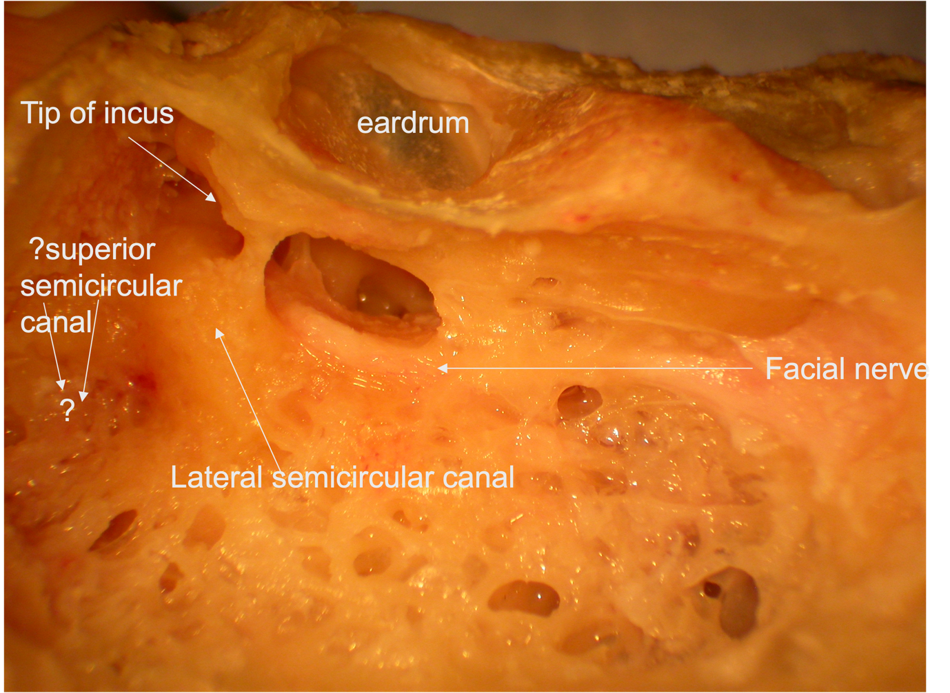 | This picture shows some of the structures that we wish to avoid, in this case deliberately exposed. Also, there might be structures like the superior semicircular canal (a canal of balance) deeply buried in bone that we need to find and operate on. |
 | This illustration shows the canal's presumed location and orientation, which we would aim to calculate by aligning the optical image with the pre-operatve CT scan. |