
|
Department of Engineering |
Cochlear implants (CI) enable deaf patients to acquire 'electronic hearing', through the insertion of an electrode array into the fluid filled cochlea within the inner ear. The surgical insertion of the CI is a potentially hazardous procedure. The CI is fed slowly through the inner ear's round window and must coil inside the cochlea. This requires reaction forces between the CI and the cochlear wall, with the potential for scarring, inflammation and (in the worst case) penetration of the basilar membrane which separates the two fluid filled scalae inside the cochlea. All of these adverse outcomes are detrimental to electronic hearing.
This project seeks to better understand the mechanics of CI insertion. Our collaborators at Addenbrooke's Hospital have developed an experimental protocol that allows robotic insertion of the implant into a transparent, 3D-printed cochlea, with simultaneous measurement of the insertion force (see here for a movie). This project will complete the analysis by simultaneously tracking the CI's electrodes in 3D. The resulting 4D reconstruction (3D space plus time) will be accomplished via two orthogonal camera views of the cochlea during insertion. Prior knowledge of the CI's geometry will be exploited to improve the accuracy of the reconstruction.
The project is offered in collaboration with Professor Manohar Bance, Dr Sita Clark and Dr Iwan Roberts at Addenbrooke's Hospital. The project would suit a student who has taken Module 3G4, though this is not a strict prerequisite. It will involve programming in Python and/or C++, and the opportunity to learn more about medical image analysis, computer vision and graphics. The results of the project are expected to enhance significantly our understanding of CI insertion mechanics and how to reduce the risk of adverse outcomes.
| A single frame of the 4D reconstruction we are seeking, this example is taken from the end of the time sequence when the CI is fully inserted. In this case, the CI has pierced the basilar membrane (red) between the scala tympani (green) and the scala vestibuli (blue). The structures we are looking at are tiny: a typical cochlea measures around 8mm across and each CI electrode is around 0.5mm long. |
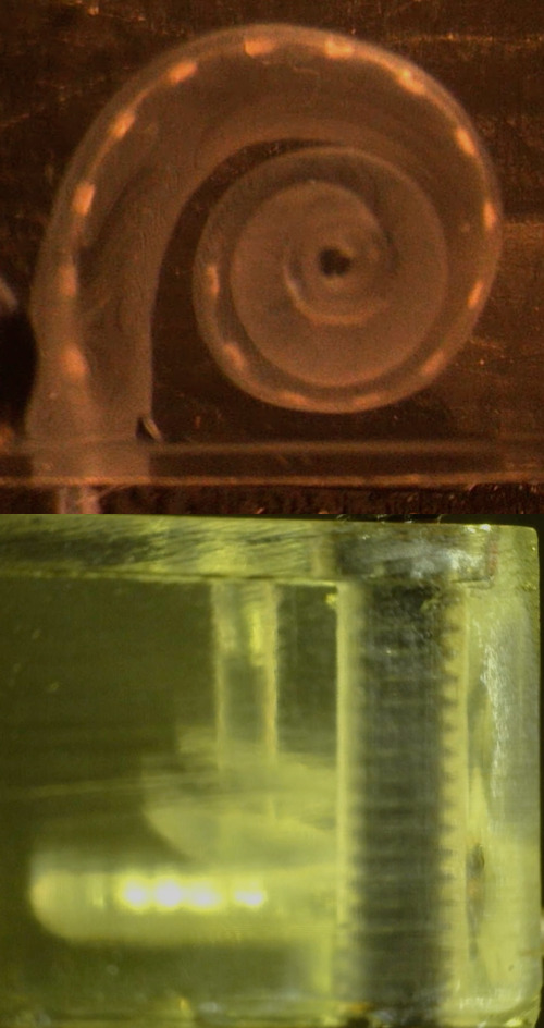 | These are single, orthogonal frames from a movie recorded during insertion of a CI into a transparent, 3D-printed cochlea. The first stage of the project will involve camera calibration, the process of determining the position and orientation of each camera with respect to the cochlea. Once calibrated, it should be possible to backproject the image of each electrode into 3D space and then fit the known geometrical model of the CI to the backprojection. The final step will be to track the 3D reconstruction in time, producing a 4D animation. |