 |
 |
Department of Engineering |
|
F-AHG-2: Simulation of middle and inner ear surgery
Ear surgery, such as cochlear implantation but other surgeries also, involves drilling through part of the skull in order to access the middle ear. This is a technically challenging procedure requiring years of training, since there are many potential hazards. These include not drilling accidently into the facial nerve (which moves the face), the inner ear (causes deafness and balance loss if hit), the sigmoid sinus (copious bleeding) or the dura (causes brain fluid leakage), to name but a few. Structures are packed together tightly and covered in bone, so not visible until the bone is drilled away. This project aims to help the surgeon practice the procedure by way of a computer graphics simulation.
Starting with a high resolution, micro-CT scan of the relevant anatomy, we will segment a triangulated mesh of the bone using shape-based interpolation and regularised marching tetrahedra, as in the 3G4 laboratory. We will then develop an interactive application that produces a real-time surface rendering of the mesh, with the viewpoint controlled by the user, analogous to the surgeon choosing where to position the surgical microscope. The main challenge will come with simulated drilling of the bone using a virtual surgical drill. This will require mastering some challenging concepts in computational geometry, so as to edit the mesh in real time as sections of bone are drilled away.
All of this has been done before in a number of virtual reality surgical simulators, for example Visible Ear Simulator, Voxel-Man and CardinalSim, though these have been decades in the making! The aim of this project is to reproduce a small section of this work, since it will provide the interested student with the perfect opportunity to learn about computer graphics and computational geometry.
The project is offered in collaboration with Professor Manohar Bance, Dr Sita Clark and Dr Iwan Roberts at Addenbrooke's Hospital. The project would suit a student with a keen interest in computer graphics who has taken Module 3G4 and Project GG2, though neither is a strict prerequisite. It will involve programming in a graphics framework of the student's choice, either low-level (OpenGL/Vulkan) or perhaps a high-level engine like Unreal.
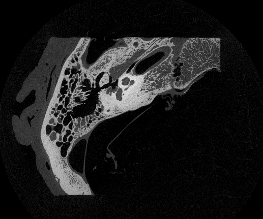 |
The starting point for the project will be high resolution micro-CT scans of the temporal bone. This image shows a slice through such a 3D volume. |
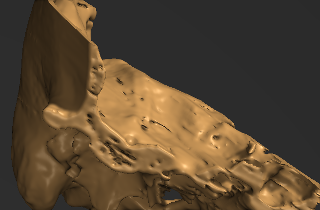 |
Producing a high quality surface mesh of the bone is not trivial. This image shows a quick proof-of-concept using thresholding and regularised marching tetrahedra. |
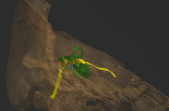 |
In virtual reality, it is possible to change the opacity of the mesh to reveal segmented internal structures, in this example the facial nerve, otic capsule and ossicles. |
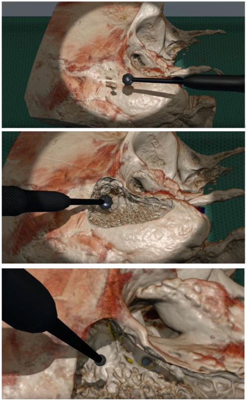 |
These pictures, reproduced from this paper, shows the CardinalSim surgical simulator. The most challenging part of the project will be devising computational geometry algorithms for real-time mesh editing as sections of bone are virtually drilled away. |