Probe calib
Stradwin incorporates automatic spatial and temporal calibration systems for
3D ultrasound, derived from the successful Stradx system. The calibration
process takes only minutes to perform.
In order to perform probe calibration, you need to do the following things:
- Fill a flat-bottomed water bath with water. The water should be
at room temperature. Ideally the temperature of the water should be
fairly constant during the calibration process.
- Mount your position sensor rigidly on the ultrasound probe. A Polaris or
Bird sensor may usually be mounted close to the body of the probe, but a
Fastrak should be fixed about 10 cm away using some sort of rigid
plastic strut.
- If you have a Cambridge phantom available, mount the probe in the
phantom, ensuring that the brass strip in the phantom is at all times
in the centre of the ultrasound beam. If you don't have a Cambridge
phantom, ensure that the bottom of the water bath is flat and roughen
it uniformly using small circular motions with very fine emery cloth
or sand paper. This will improve the reflection of ultrasound at
oblique angles.
- Check that you have Stradwin running with both images and
positions available. This information is displayed on the 'Record'
task page. The configuration dialogs for both images and positions
can also be accessed from this page. Use the
'image source' dialog to select the
image source you wish to use and the area of the ultrasound scanner
screen that you want to record. When you are happy with the image
cropping you should save it to a template file using the
file menu, so you can use it with other
calibrations if required.
- On the 'Record' task page, set the 'Video Rate' to 'Full Speed'.
After changing this value, select the 'Probe Calib' task page.
- Switch on live display mode using the (leftmost) button on the
toolbar. Configure the controls of the ultrasound machine so that the
line feature appears clearly in the B-scan image.
- If you have a curvilinear probe, use the 'Define region of interest'
button to outline the region of the image occupied by the scan. The
left mouse button is used to specify each point round the edge of the
region, and the right button is used to indicate the final point.
- Set the controls of the ultrasound machine so that the line
detector locks on to the bottom of the water bath (or bar of the
Cambridge phantom).
- Select the 'Probe Calib' task page and press the 'Toggle temporal
calibration' button. Stradwin is now in temporal calibration mode. Follow the
instructions at the top of the task page. You will be asked to hold the probe
steady while imaging the bottom of the water bath. You will then be asked to
move the probe up and down while Stradwin tracks the bottom of the water bath
in the image. By matching the image motion with the readings from the position
sensor, Stradwin can estimate the temporal calibration, which is the relative lag
between the position and image streams. The result is presented in terms of the
lag, and also a confidence value, representing how how well the image and
position streams correlated. You should repeat the temporal calibration a few
times (by pressing the 'Toggle temporal calibration' button again), and obtain
a few results with confidences exceeding 95%. Keep repeating this process until Stradwin
displays a temporal calibration result that is in line with the
>95% confidence consensus, then move on to the next step.
- Measure the temperature of the water in Celsius (degrees
Centigrade), enter the value in the box on the 'Probe Calib' task page
and press the 'Set' button. Note that the temperature compensation strategy is
not completely effective for probes with very high curvature, such as rectal
probes. For these probes, it will be more accurate to calibrate with warm water
as close as possible to 48 Celsius.
- This next step involves estimating the x and y image scales (mm per
pixel). You should skip this step if you have an RF image source, or are using a Terason or Sonix, in which case the scales are implicit. Most ultrasound machines have facilities for measuring distances
in the image. Use a facility of this sort to display either a line of
a known length, or two marks a known distance apart. You need to be
able to see both marks, or both ends of the line, in the live Stradwin
image window. For maximum accuracy, the line should not be short: it should
occupy a good portion of the visible ultrasound image. Click the button 'Mark
ends of first line', and click the left mouse button on one end of the line and
the right mouse button on the other end. Then click the 'Finished locating ends of line'
button. Set the slider as close as you can to the correct length of
the line in millimetres. Repeat this process for a second line (use the 'Mark
ends of second line' button), which should be roughly perpendicular to the first line.
- Go through the sequence of movements listed below
and, when you are happy that the line detector is locked onto the
bottom of the water bath in each position, click the 'Accept line for
calibration' button. If the line detector is not happy with the line
it will display 'No valid line' on this button, but you also need to
exercise judgement to avoid capturing incorrect lines. If you
mistakenly accept a line that is not an image of the bottom of the
bath (or bar of the Cambridge phantom), then you should click the 'Delete all
accepted lines' button and start again.
- You should aim to acquire about 40 lines, as described below.
- Press the 'Solve for spatial calibration' button to run the
calibration solution algorithm. This algorithm uses the recorded
positions of those lines which were accepted.
- Enter the name of the ultrasound probe and the depth setting in the two
boxes. Since a calibration applies only to a specific probe and depth setting,
it is very important that you correctly identify these at this stage. You need
to press 'enter' in both boxes before the 'Accept calibration' button is
enabled.
- Press the 'Accept calibration' button. The effect of the new calibration
is now apparent in all of Stradwin's windows. However, the calibration is not
yet saved to file.
- On the 'Record' task page, set the 'Video rate' to the
value that you will require when you reload the template to
use it for recording data. The recommended value for this
is the 'Gated' setting.
- Save the calibration to a template file using the file menu.
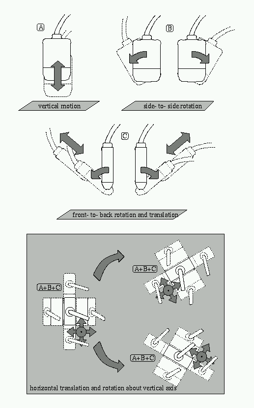
Motion sequence for calibration
In what follows, the term 'phantom plane' refers to the bottom of the water
bath or the virtual plane swept out by the bar of the Cambridge phantom.
Initially position the probe above the phantom plane. This is the
starting position for all the movements described below.
(A) Move the probe vertically up and down without changing its
orientation.
(B) Rotate the probe from side to side while keeping the scan plane
and the phantom plane perpendicular. You should rotate the probe both
clockwise and anticlockwise from its starting position.
(C) Rotate the probe towards you and away from you, changing the
angle between the scan plane and the phantom plane.
Note that the angle of the image of the phantom plane
should not change significantly during these moves.
(D) Rotate the probe through 45 degrees in each direction about its
vertical axis.
(E) Translate the probe across the phantom plane and perform motions
A, B, C and D in a total of three non-collinear locations.
You should aim to acquire about 40 lines.
Verification of calibration consistency with the pointer
Wherever possible, you should evaluate your calibration by testing
whether or not it is consistent with your pointer calibration.
To do this you need to have a Polaris position sensor that is able to track
two things simultaneously.
Ensure that the tracked probe and a trackable pointer
are both in the active volume of the position sensor.
Use the
position source dialog
from the
configuration menu
to get Stradwin to track both of them. You should be able
to see them both in the 3D window when live display is enabled
(using the left-most button on the toolbar).
If the pointer has not already been calibrated, calibrate
it as described on the pointer calibration page.
Ensure that the pointer calibration task
page is selected. While keeping the probe in the tracked volume,
dip it in a water-bath and hold the tip of the pointer in the beam.
You should be able to see the reflections from the tip of the pointer
in the live B-scan image. If the calibration of the probe and the
pointer are consistent, you will also see a dot with a circle around
it. The dot indicates the location of the pointer in the B-scan
calculated by the system. The radius of the circle indicates the
measured distance of the pointer tip from the plane of the B-scan.
If the calibrations are consistent, the circle should be small when the
pointer tip shows up most strongly in the B-scan image. Note that
there is a small delay in displaying the pointer position in the
image. This is not a sign of a problem with the system.
The goal is to ensure consistency when both the pointer and the
ultrasound probe are effectively stationary.
It is important to repeat the consistency check described in the
previous paragraph with the pointer tip at various different positions
in the B-scan image. You should make sure you check out all parts of the
B-scan, not just the centre or one corner.
Controls affecting the line-fitting algorithm
- Variance of Gaussian kernel
- This controls the smoothing applied to the image
in advance of the edge detection process.
- Vertical analysis bands
- This sets the number of vertical bands in the image
that are used for the edge detection process. They are indicated
in the image window by vertical red lines drawn down the
image.
- Gradient threshold
- This threshold governs the way the line detection algorithm
detects edge elements. The algorithm scans down the image and picks the
first local maximum which exceeds the threshold, or the biggest local
maximum if none exceed the threshold.
- Pixel linearity threshold
- The random sample consensus (Ransac) algorithm requires a minimum
proportion of the edge elements to be collinear, otherwise
the line is not considered reliable. This parameter determines the
vertical tolerance (in pixels) that is used when determining collinearity.
- Ransac acceptance proportion
- This is the proportion of edge elements that must be collinear for
the line to be accepted.
- Configure for upside down image
- It is conventional with some probes (eg. endovaginal) to view the
image upside down, so that the probe face is at the bottom of the
image and objects distant from the probe at the top of the image. By clicking
on the 'Configure for upside down image' button, the edge detector will scan up
from the bottom of the image for the
first strong edge, instead of down from the top. Click this button
when the image is inverted.