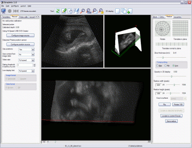
This page gives a selection of views of Stradwin being used in a variety of applications. Click on an image to see a larger version.
Reslice of a foetus
Polaris volumes in the 3D window, probe calibration verification in the image window.
Structure of a talipes foot in 3D
Orthogonal slices of a bladder
Plaque near a blood vessel bifurcation
Strain image of a phantom containing half an olive
Example of a data set requiring probe pressure correction
Panoramic image of a both lobes of a human thyroid
Multiple sweep data covering an entire human liver
Reconstructed steered RF data and raw RF visualisation
3D RF data from a convex 3D probe
Cortical thickness estimation from CT data
CT data from a DICOM file