Difference: GMT_4YP_17_1 (r2 vs. r1)
| r2 - 24 Mar 2017 - 12:51 - Main.gmt11 | r1 - 24 Mar 2017 - 11:50 - Main.gmt11 | ||||||||||||||||||||||||||||||||||||||||||||||
|---|---|---|---|---|---|---|---|---|---|---|---|---|---|---|---|---|---|---|---|---|---|---|---|---|---|---|---|---|---|---|---|---|---|---|---|---|---|---|---|---|---|---|---|---|---|---|---|
Dr Graham Treece, Department of Engineering |
Dr Graham Treece, Department of Engineering |
||||||||||||||||||||||||||||||||||||||||||||||
F-GMT11-1: Measuring bone cortical thickness of the skull |
F-GMT11-1: Measuring bone cortical thickness of the skull |
||||||||||||||||||||||||||||||||||||||||||||||
|
|
||||||||||||||||||||||||||||||||||||||||||||||
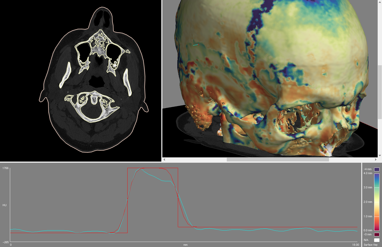
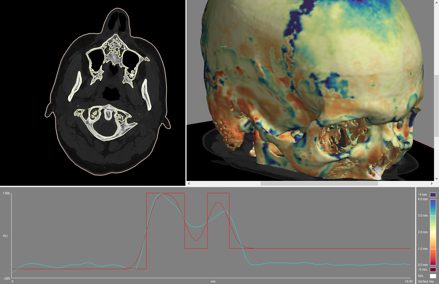
Bone fracture is a major issue affecting millions of people annually, and we have recently been involved in research which has the potential to contribute significantly to both our understanding of fracture and how various preventative measures affect bone. The advances have come from a much more precise (and hence much more sensitive) measurement of the bone cortex (the denser layer surrounding the less dense bone in the middle). As well as contributing to our understanding of fracture, this technique is also being used to underpin models of bone used in mechanical analysis, for instance to see what happens to the skull during a head injury. It is also increasingly being used by paleo-anthropologists who are interested in the properties of very old bones. |
Bone fracture is a major issue affecting millions of people annually, and we have recently been involved in research which has the potential to contribute significantly to both our understanding of fracture and how various preventative measures affect bone. The advances have come from a much more precise (and hence much more sensitive) measurement of the bone cortex (the denser layer surrounding the less dense bone in the middle). As well as contributing to our understanding of fracture, this technique is also being used to underpin models of bone used in mechanical analysis, for instance to see what happens to the skull during a head injury. It is also increasingly being used by paleo-anthropologists who are interested in the properties of very old bones. |
||||||||||||||||||||||||||||||||||||||||||||||
The existing technique expects to measure a single 'layer' of bone, but in many places bone has more than one layer: for instance in the skull there are two layers: an inner and an outer table, which in some places join together to become just one. In these areas it is not clear whether our measurements relate to one or both tables. The aim of this project is to try to disentangle this information, by looking at measurements made from either side of the bone, to try to split them into clean sets of measurements of either the whole skull thickness, or just the inner and outer tables. This is an interesting problem in computational geometry, but also in surface-based registration and visualisation |
The existing technique expects to measure a single 'layer' of bone, but in many places bone has more than one layer: for instance in the skull there are two layers: an inner and an outer table, which in some places join together to become just one. In these areas it is not clear whether our measurements relate to one or both tables. The aim of this project is to try to disentangle this information, by looking at measurements made from either side of the bone, to try to split them into clean sets of measurements of either the whole skull thickness, or just the inner and outer tables. This is an interesting problem in computational geometry, but also in surface-based registration and visualisation |
||||||||||||||||||||||||||||||||||||||||||||||
This is an algorithmic development / computational geometry / software project, so experience of writing software is essential, though the development could also be done using Matlab. |
This is an algorithmic development / computational geometry / software project, so experience of writing software is essential, though the development could also be done using Matlab. |
||||||||||||||||||||||||||||||||||||||||||||||
Click here for other medical imaging projects offered by Graham Treece. |
Click here for other medical imaging projects offered by Graham Treece. |
||||||||||||||||||||||||||||||||||||||||||||||
|
|
||||||||||||||||||||||||||||||||||||||||||||||
| r2 - 24 Mar 2017 - 12:51 - Main.gmt11 | r1 - 24 Mar 2017 - 11:50 - Main.gmt11 | ||||||||||||||||||||||||||||||||||||||||||||||

