Difference: GMT_4YP_18_3 (r4 vs. r3)
| r4 - 26 Apr 2021 - 19:42 - Main.gmt11 | r3 - 21 Apr 2021 - 09:01 - Main.gmt11 | ||||||||||||||||||||||||||||||||||||||||||||||||||||||||||||||||||||||||||||||||||||||||||||||||||||||||||||||||||||
|---|---|---|---|---|---|---|---|---|---|---|---|---|---|---|---|---|---|---|---|---|---|---|---|---|---|---|---|---|---|---|---|---|---|---|---|---|---|---|---|---|---|---|---|---|---|---|---|---|---|---|---|---|---|---|---|---|---|---|---|---|---|---|---|---|---|---|---|---|---|---|---|---|---|---|---|---|---|---|---|---|---|---|---|---|---|---|---|---|---|---|---|---|---|---|---|---|---|---|---|---|---|---|---|---|---|---|---|---|---|---|---|---|---|---|---|---|---|
Dr Graham Treece, Department of Engineering |
Dr Graham Treece, Department of Engineering |
||||||||||||||||||||||||||||||||||||||||||||||||||||||||||||||||||||||||||||||||||||||||||||||||||||||||||||||||||||
F-GMT11-3: Reducing noise due to metal in CT data |
F-GMT11-3: Reducing noise due to metal in CT data |
||||||||||||||||||||||||||||||||||||||||||||||||||||||||||||||||||||||||||||||||||||||||||||||||||||||||||||||||||||
|
|
||||||||||||||||||||||||||||||||||||||||||||||||||||||||||||||||||||||||||||||||||||||||||||||||||||||||||||||||||||
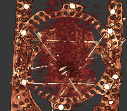
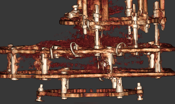
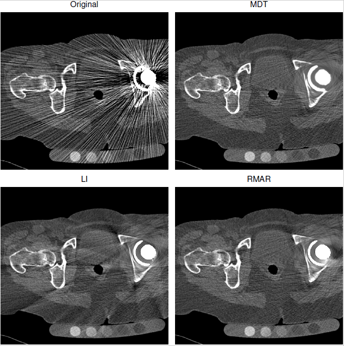
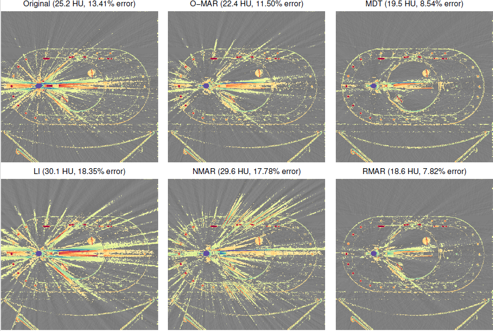
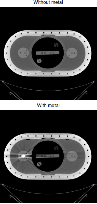
Metal in CT data is very difficult to image, because the metal is so dense compared to bone or tissue, and hence doesn't let many of the X-rays through. This causes problems when the data is reconstructed (i.e. the cross-sectional image re-created from the measured projections). The assumptions made in the reconstruction algorithm aren't valid for this situation, and the result is errors over the whole reconstructed image, often appearing as radial lines emanating from the metal. But CT imaging is used for all sorts of things in medicine: and many people have some sort of metal in their body, either from a hip replacement, or stent, or even from a filling. For instance, this presents a major problem for orthopaedic surgeons who typically would use CT scans to provide the best imaging data for patient treatment. |
Metal in CT data is very difficult to image, because the metal is so dense compared to bone or tissue, and hence doesn't let many of the X-rays through. This causes problems when the data is reconstructed (i.e. the cross-sectional image re-created from the measured projections). The assumptions made in the reconstruction algorithm aren't valid for this situation, and the result is errors over the whole reconstructed image, often appearing as radial lines emanating from the metal. But CT imaging is used for all sorts of things in medicine: and many people have some sort of metal in their body, either from a hip replacement, or stent, or even from a filling. For instance, this presents a major problem for orthopaedic surgeons who typically would use CT scan to provide the best imaging data for patient treatment. |
||||||||||||||||||||||||||||||||||||||||||||||||||||||||||||||||||||||||||||||||||||||||||||||||||||||||||||||||||||
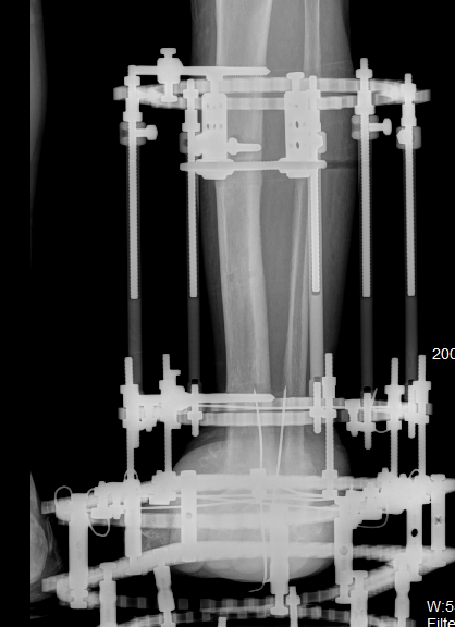
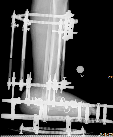
Most of the correction techniques look at the original projections (the actual data recorded by the CT scanner), work out which bits of these projections were affected by metal, and then try to make up new projection data to replace the corrupted data. There are various techniques for this, among which Refined Metal Artefact Reduction (RMAR), developed at Cambridge, works particularly well. However, it presumes an X-ray geometry which is reasonably planar. Hence it works well for standard helical CT, but less well for cone beam CT, which is increasingly being used for instance in knee and ankle scanning. |
Most of the correction techniques look at the original projections (the actual data recorded by the CT scanner), work out which bits of these projections were affected by metal, and then try to make up new projection data to replace the corrupted data. There are various techniques for this, among which Refined Metal Artefact Reduction (RMAR), developed at Cambridge, works particularly well. However, it presumes an X-ray geometry which is reasonably planar. Hence it works well for standard helical CT, but less well for for cone beam CT, which is increasingly being used for instance in knee and ankle scanning. |
||||||||||||||||||||||||||||||||||||||||||||||||||||||||||||||||||||||||||||||||||||||||||||||||||||||||||||||||||||
|
|
||||||||||||||||||||||||||||||||||||||||||||||||||||||||||||||||||||||||||||||||||||||||||||||||||||||||||||||||||||
The aim of this project is to try to adapt RMAR for the very non-planar geometry in cone beam CT and, in the process, hopefully also improve it for the slightly non-planar geometry in more conventional helical CT. Our orthopaedic surgery partners at the Paley Institute Florida USA have provided us with patient imaging and are also available for clinical relevance guidance. Successful artefact reduction would enable the better assessment of bone healing. |
The aim of this project is to try to adapt RMAR for the very non-planar geometry in cone beam CT and, in the process, hopefully also improve if for the slightly non-planar geometry in more conventional helical CT. Our orthopaedic surgery partners at the Paley Institute Florida USA have provided us with patient imaging and are also available for clinical relevance guidance. Successful artefact reduction would enable the better assessment of bone healing. |
||||||||||||||||||||||||||||||||||||||||||||||||||||||||||||||||||||||||||||||||||||||||||||||||||||||||||||||||||||
This is an algorithmic development / software project, so experience of writing software is essential, though the development could also be done using Matlab. It would ideally suit someone with experience of both the 3G4 medical imaging and the GG2 CT reconstruction IIA project, or at least one of these. |
This is an algorithmic development / software project, so experience of writing software is essential, though the development could also be done using Matlab. It would ideally suit someone with experience of both the 3G4 medical imaging and the GG2 CT reconstruction IIA project, or at least one of these. |
||||||||||||||||||||||||||||||||||||||||||||||||||||||||||||||||||||||||||||||||||||||||||||||||||||||||||||||||||||
Click here for other medical imaging projects offered by Graham Treece. |
Click here for other medical imaging projects offered by Graham Treece. |
||||||||||||||||||||||||||||||||||||||||||||||||||||||||||||||||||||||||||||||||||||||||||||||||||||||||||||||||||||
|
|
||||||||||||||||||||||||||||||||||||||||||||||||||||||||||||||||||||||||||||||||||||||||||||||||||||||||||||||||||||
| r4 - 26 Apr 2021 - 19:42 - Main.gmt11 | r3 - 21 Apr 2021 - 09:01 - Main.gmt11 | ||||||||||||||||||||||||||||||||||||||||||||||||||||||||||||||||||||||||||||||||||||||||||||||||||||||||||||||||||||


