Difference: GMT_4YP_18_3 (r2 vs. r1)
Dr Graham Treece, Department of Engineering
F-GMT11-3: Reducing noise due to metal in CT data
 |  |  |
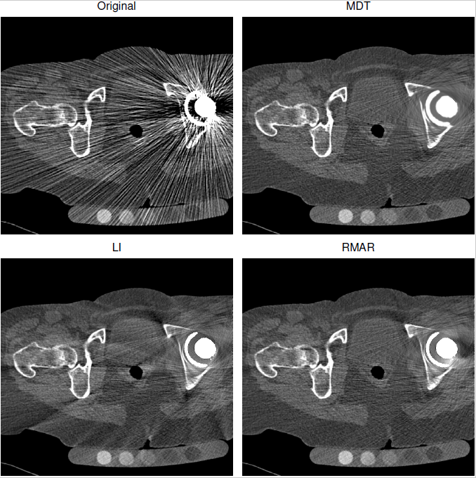
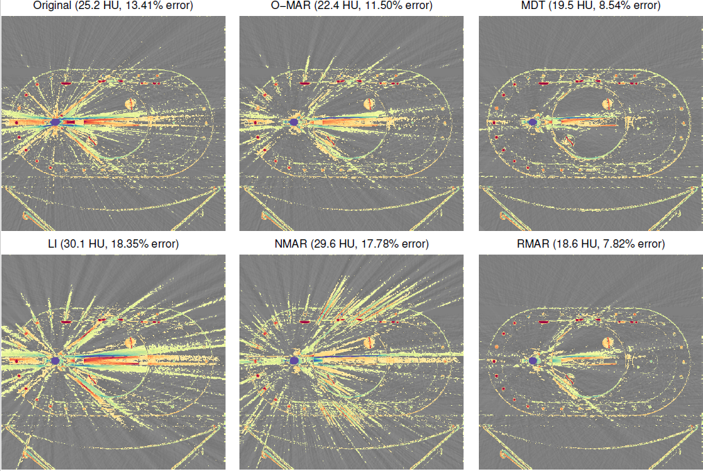
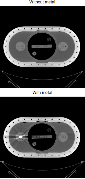
| Here are some typical CT images of someone with a metal hip replacement. Top left shows the errors that result from having metal in the CT scans, and the other images are various forms of correction for these artefacts. Each of these corrections is based on replacing the projected data which has been affected by metal. | It is easier to assess this when scanning a known object: above is a phantom without any metal in, and below scanned again after inserting a pretend hip replacement. | The colour in these images is the difference between the corrected scan, and the real scan with no metal in it, so colours show errors which were not corrected in each case. All of the corrections still have errors over the image a long way from the actual metal, because it is really hard to know how to 'fill in' the gap in the projections where the metal was. |
Metal in CT data is very difficult to image, because the metal is so dense compared to bone or tissue, and hence doesn't let many of the X-rays through. This causes problems when the data is reconstructed (i.e. the cross-sectional image re-created from the measured projections). The assumptions made in the reconstruction algorithm aren't valid for this situation, and the result is errors over the whole reconstructed image, often appearing as radial lines emanating from the metal. But CT imaging is used for all sorts of things in medicine: and many people have some sort of metal in their body, either from a hip replacement, or stent, or even from a filling. So the correction of these errors is an important topic.
Most of the correction techniques look at the original projections (the actual data recorded by the CT scanner), work out which bits of these projections were affected by metal, and then try to make up new projection data to replace the corrupted data. However, the form of this new projection data has a significant impact on the final reconstructed images: simply assuming a straight line between surrounding data, for instance, creates significant new errors. A better guess would be to use the reconstructed data to help replace the projected data: but the reconstructed data is corrupted by the errors we are trying to remove ... hence this project will investigate different ways of trying to guess the form of the missing projection data, without simply reintroducing the metal errors.
This is an algorithmic development / software project, so experience of writing software is essential, though the development could also be done using Matlab. It would ideally suit someone with experience of both the 3G4 medical imaging and the GG2 CT reconstruction IIA project, or at least one of these.
Click here for other medical imaging projects offered by Graham Treece.
| I | Attachment | Action | Size | Date | Who | Comment |
|---|---|---|---|---|---|---|
| ct29_frame135_1.png | manage | 396.7 K | 23 Mar 2018 - 12:00 | UnknownUser | ||
| data3_frame99_1.png | manage | 747.0 K | 23 Mar 2018 - 12:00 | UnknownUser | ||
| data3_frame99_2.png | manage | 90.2 K | 23 Mar 2018 - 12:00 | UnknownUser |