Difference: GMT_4YP_21_2 (r2 vs. r1)
Dr Graham Treece, Department of Engineering
F-GMT11-2: Ankle prosthesis alignment from X-rays
  |   |   |
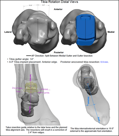
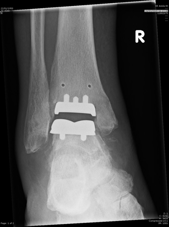
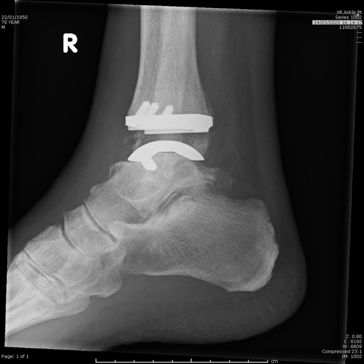
| An image The implant location on the tibia and the talus is planned based on a pre-operative CT scan of the patient | An image A post-operative CT isn't usually available, but X-ray images are | An image Can the implant axial rotation with respect to the tibia be calculated from these images? |
A description.
Click here for other medical imaging projects offered by Graham Treece.
| I | Attachment | Action | Size | Date | Who | Comment |
|---|---|---|---|---|---|---|
| Picture1.png | manage | 131.2 K | 13 Apr 2021 - 10:01 | UnknownUser | ||
| Picture2.png | manage | 328.5 K | 13 Apr 2021 - 10:01 | UnknownUser | ||
| Picture3.png | manage | 387.2 K | 13 Apr 2021 - 10:01 | UnknownUser |
View topic | View difference side by side | History: r4 < r3 < r2 < r1 | More topic actions
No permission to view System.WebBottomBarExample