Strain imaging in Stradwin
Funded by the Wellcome Trust, this technology transfer project aims to develop two-dimensional (2D) ultrasonic elasticity imaging techniques to a stage where they can be licensed to manufacturers of ultrasound equipment. Part of this process involves clinical trials at Addenbrooke's Hospital, with clinical feedback informing algorithmic and ergonomic refinement.
This project addresses an unmet clinical need for reliable, cost-effective, non-invasive characterisation of stiff lesions. Stiffness is of course an important indicator of disease. Ultrasonic elasticity imaging meets this clinical need by offering quantitative measurements of tissue strain several centimetres below the skin surface.
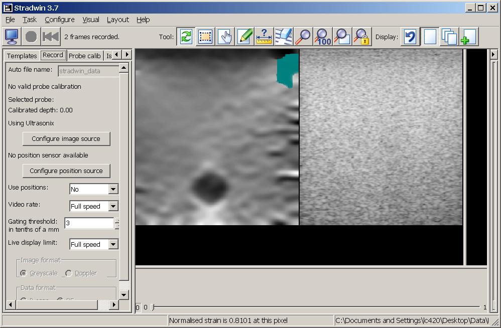
The broad objective of this project is to develop a market-ready ultrasonic elasticity imaging system, capable of producing reliable images of tissue strain in a clinical environment. Such a system would find widespread application in oncology and other areas. We adopt a simple approach whereby the clinician gently compresses the tissue with the ultrasound probe during the examination. In the literature, this is referred to as “freehand quasistatic compression” elasticity imaging. It has the benefit of working with all existing ultrasound scanners: there is no need for any hardware modification. By modifying the scanner’s software, we can arrange for the machine to display strain images instead of the usual backscatter images. An example is shown in Fig. 1, in which a strain image and a B-mode image is displayed side-by-side in Stradwin.
|
|
|
Strain imaging in Stradwin |
We have novel algorithms for strain imaging which offer world leading performance both in terms of signal-to-noise ratio and speed. We also have considerable experience in 3D ultrasonic imaging which we have recently applied to 3D elasticity imaging. The goals of this project are to take this technology out of the laboratory and develop it into a state where it can be licensed to manufacturers of ultrasound equipment. The performance of the software will be established through clinical studies documented in peer-reviewed academic publications.
In the course of this research project, we have created a clinical database of ultrasonic radio frequency (RF) strain imaging data. The database aims to assist evaluating our novel strain imaging algorithms and it also provides the elastography research community a valuable clinical resource.
Approval was granted by the Cambridgeshire−3 Research Ethics Committee for the anonymized data to be disseminated to the wider research community. Parties interested in accessing the data should send a message to stradwin-info@eng.cam.ac.uk .
|
|
|
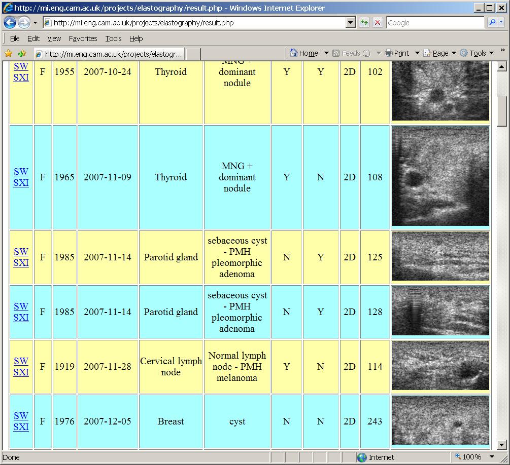
Web interface of the clinical database |
Click here to access the database.
Based on several data-processing methods, including a hybrid discrimination estimation algorithm [1, 3], a novel data-weighting scheme and a noise-masking approach [2], developed in this project, we have identified many cases that demonstrates the potential clinical application of strain imaging [2]. The following examples serve to illustrate the distinctive features of the strain images we produce and how these may help clinicians to make better diagnosis.
|
|
|
|
|
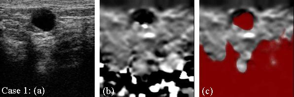
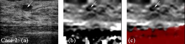
The B-mode images (a) show small hypo/anechoic breast lesions with marked posterior acoustic enhancement. Differentiation between cysts and solid benign fibroadenomata is not always straightforward. The corresponding strain images confirm stiff lesions in both instances, but the colour wash masks the fluid within the cyst wall (case 1). There is no colour wash over the fibroadenoma (indicated in case 2), which retains the expected characteristics of stiffness in the strain image. Scatterer motion is not coherent between pre- and post-deformation frames in lesions containing fluid. The colour wash is therefore able to distinguish between cystic and solid lesions. |
The clinical relevance of ultrasonic strain imaging has yet to be established. In a recent evidence based review (Cole JA, et al. Evidence review, Ultrasound elastography. NHS Purchasing and Supply Agency, Centre for Evidence-based Purchasing; 2009. CEP08052), only transient ultrasound elastography (a quantitative technique for making point stiffness measurements, not images) emerged as a clinically proven technique for assessing liver fibrosis. For breast imaging, the conclusion was that ultrasound elastography “may prove to be a useful addition . . . perhaps serving as an adjunct to conventional ultrasound, rather than a replacement for it, but further research is needed.” Only “limited evidence” was found for ultrasound elastography’s utility in prostate, endoscopic and vascular imaging. It is therefore still early days for this technology, but the commitment of the major equipment manufacturers suggests an expectation that its utility will eventually be proven.
L. Chen, R. J. Housden, G. M. Treece, A. H. Gee, and R. W. Prager, “A hybrid displacement estimation method for ultrasonic elasticity imaging”, To appear in IEEE Transactions on Ultrasonics, Ferroelectrics, and Frequency Control.
L. Chen, S. J. Freeman, A. H. Gee, R. J. Housden, R. W. Prager, R. Sinnatamby, G. M. Treece, “Initial clinical experience of an ultrasonic strain imaging system with novel noise-masking capability”, The British Journal of Radiology.
L. Chen, G. M. Treece, J. E. Lindop, A. H. Gee, and R. W. Prager, “A quality-guided displacement tracking algorithm for ultrasonic elasticity imaging”, Medical Image Analysis.
A. H. Gee, L. Chen, S. Freeman, G. M. Treece, R. W. Prager, and L. H. Berman, “A clinical database for evaluating freehand quasistatic strain imaging systems”, Seventh International Conference on the Ultrasonic Measurement and Imaging of Tissue Elasticity, Austin, Texas, USA.
|
© Department
of Engineering at the University of Cambridge |
Last updated: January 2009