|
|
|||
 |
Department of Engineering |
| University of Cambridge > Engineering Department > Machine Intelligence Lab > Medical Imaging Group |
High Definition Three-Dimensional Ultrasound
EPSRC Grant GR/N21062
The objectives of this project are:
Freehand 3D ultrasound is acquired by attaching a position sensor to the probe of a conventional 2D diagnostic ultrasound machine. As the clinician moves the probe, its position and orientation are recorded with respect to some fixed datum. The 2D ultrasound images are also recorded. These 2D slices, together with the information about their positions and orientations, constitute an irregularly sampled 3D dataset describing the volume scanned by the clinician.
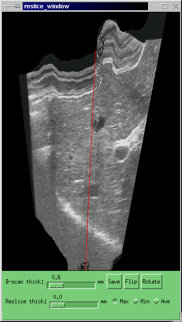
Using 3D ultrasound, clinicians can view slices through the body that would be inaccessible using a normal probe. Curved reslices can be generated to help in the early diagnosis of particular conditions such as spinabifida. The volumes of organs, glands and other structures can be calculated with much greater accuracy than is possible using a set of independent 2D measurements. This enables more accurate assessment of the progression of a disease or the response of a patient to treatment. The geometry of complex three dimensional anatomy can be visualised using 3D ultrasound, replacing X-ray CT (computed tomography) or MRI (magnetic resonance imaging) scans, which are much more expensive. X-ray CT also involves a radiation dose which is often undesirable.
Two dimensional ultrasound generally provides insufficient context for the data to be stored or transferred to a remote location for further consideration by a specialist. Three dimensional ultrasound overcomes this problem and opens the way to a range of tele-medical applications.
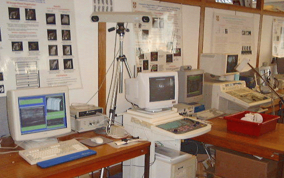
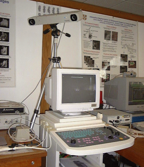
We have developed a high-resolution version of the Stradx 3D ultrasound acquisition system. Three elements of the existing design were enhanced to improve the accuracy and resolution: the 2D ultrasound images come from a Dynamic Imaging `Diasus' machine with resolution up to 0.05mm; these images are digitally transferred from the ultrasound machine to a computer; and the position sensing is performed using an optical rather than a magnetic system.
A high bandwidth ethernet link is used to transfer the digital ultrasound data from the Diasus machine to the computer running Stradx. This enables us to eliminate errors introduced by the PAL image encoding, and the A-D converter in the frame grabber.
To obtain the positions of the probe with a resolution comparable to the definition within the images, we need to use a position sensor with greater precision than the magnetic devices in our existing system. The `Polaris' system made by Northern Digital Inc. has a precision of up to 0.01mm and makes a minimal impact on the convenience with which the clinician can manipulate the ultrasound probe. Stradx now works with this system as well as with two types of magnetic position sensor.
We expect that the increase in accuracy of the optical position sensor over the magnetic system, and the improvement in the resolution of the raw 2D images, will lead to an improvement in the accuracy of our calibration algorithm. We will extend our calibration facility to enable clinicians to change the depth setting on the probe in use without repeating the complete calibration process.
All freehand 3D ultrasound systems are susceptible to serious reconstruction artefacts caused by the acquisition process itself. To maintain good acoustic contact, the clinician must hold the probe against the subject's skin with some pressure: this inevitably deforms the anatomy beneath the probe, and it is this deformed anatomy which is captured in the 3D ultrasound data set. Unless the clinician maintains perfectly constant pressure throughout the scan, the different parts of the anatomy will be captured with different degrees of compression, leading to artefact-ridden reconstructions like the ones below left. High definition scans are particularly susceptible to this kind of artefact, since the magnitude of the pressure deformation may be a significant proportion of the total image size.
We therefore correct pressure-induced artefacts using sequential image correlation techniques. A rigid in-plane translation and non-rigid depth compression are applied to each B-scan, so as to maximise the correlation between successive images. Once this has been done to all the B-scans, the 3D reconstruction will be locally smooth but may suffer from large-scale error accumulation. The entire data set is therefore subjected to one final, rigid affine transformation, so that the first and last B-scans in the sequence coincide with their externally measured positions. The image-based registration ensures that the reconstruction is locally smooth, while the position sensor readings guarantee the large-scale positional accuracy of the data.
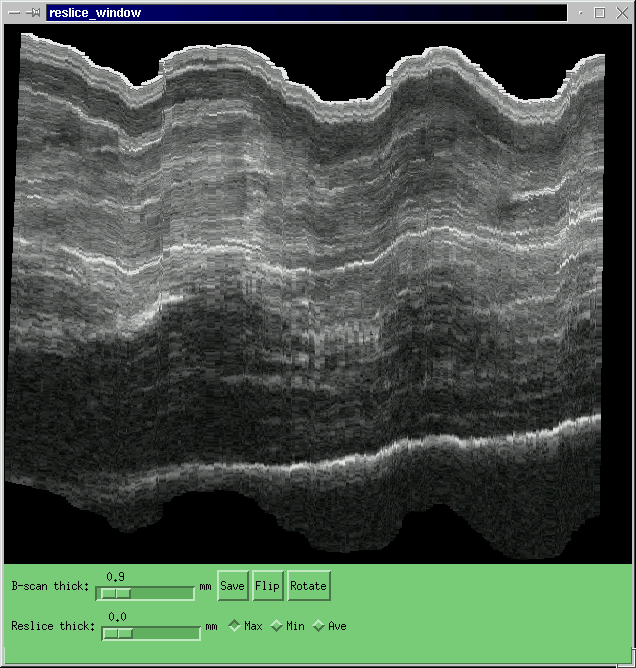
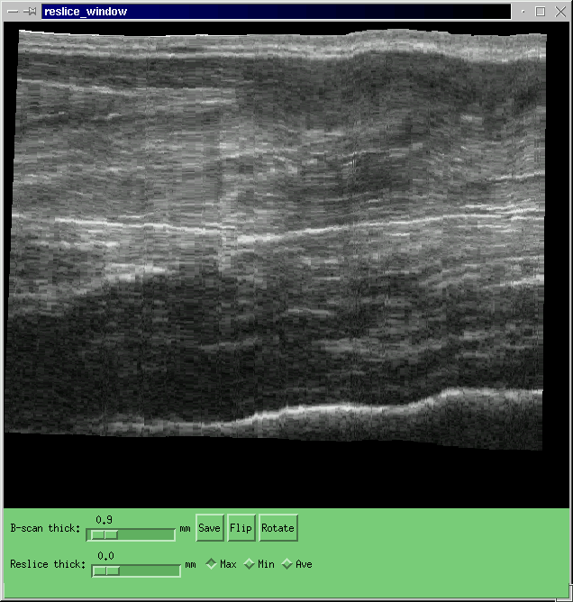
The figure above shows the original (left) and corrected (right) reslice through a 3DUS volumetric data set of the forearm.
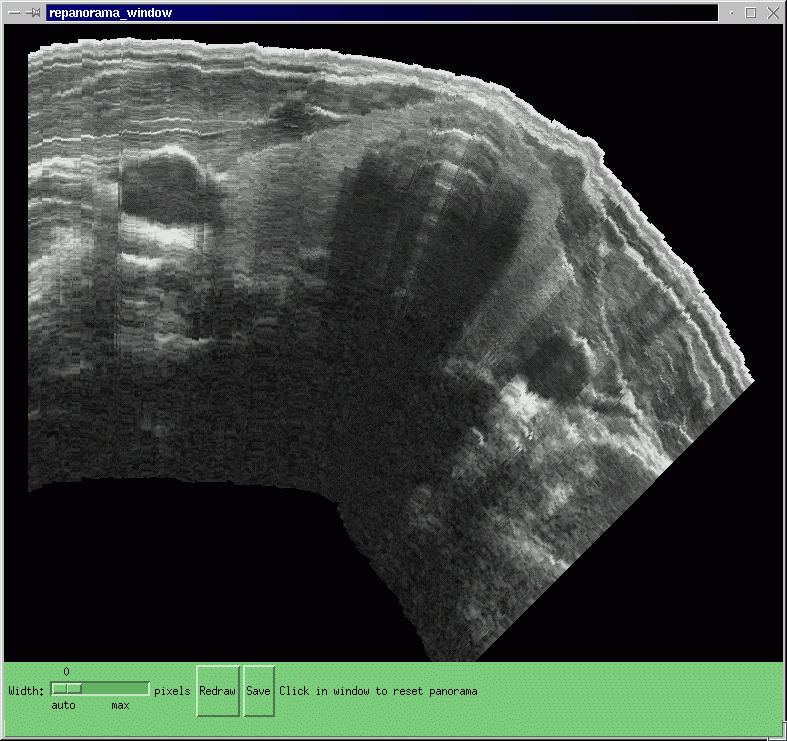

This procedure is also of benefit for panoramic data sets, for instance this of both lobes of the thyroid.
We are also investigating the application of the probe pressure correction algorithm described above to convex probes. For instance, the following figure shows one B-scan from a sequence of scans of the liver. The left hand image is the original: the right hand image has been corrected for convex probe pressure.

The effect of this correction on the whole data set can be seen in a reslice through all the original scans. Once again, the corrected image is on the right.

G. M. Treece, R. W. Prager, A. H. Gee and L. Berman. Correction of probe pressure artifacts in freehand 3D ultrasound. Accepted for oral presentation at MICCAI 2001, Utrecht, The Netherlands, October 2001.
A.H. Gee, R.W. Prager, G.M. Treece and L. Berman. Engineering a freehand 3D ultrasound system. To appear in Pattern Recognition Letters.
R.W. Prager, A.H. Gee and L. Berman. Stradx: real-time acquisition and visualization of freehand three-dimensional ultrasound. Medical Image Analysis, 3(2):129-140, 1999.
R.W. Prager, R.N. Rohling, A.H. Gee and L. Berman. Rapid calibration for 3-D freehand ultrasound. Ultrasound in Medicine and Biology, 24(6):855-869, July 1998.
A complete list of the on-line publications from our group is available here.


| | Search | CUED | Cambridge University | |
|
©
2005 Cambridge University Engineering Dept
and Graham Treece
.
Information provided by gmt11 |