Main > GMT_4YP_14_2
Dr Graham Treece, Department of Engineering
F-GMT11-2: Computer graphics: fracturing a simulated femur.
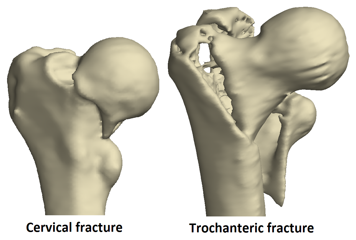
There are various reasons for wanting to study the properties of a fractured femur. But extracting the surface of a fractured femur is very complicated: here are two (very bad) examples of fractured femoral surfaces. |
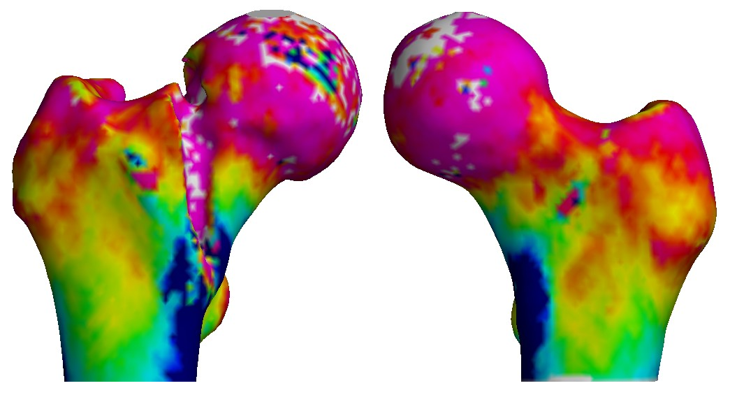
We usually also have a non-fractured femur (people don't generally facture both sides!) which is often extremely similar, other than not being fractured. Comparing this with the fractured femur is very useful: here the colour shows the thickness of the outer bone layer, which has a similar distribution on both femurs. Can we use the non-fractured femoral surface, and deliberately 'fracture' it to match the fractured side? |
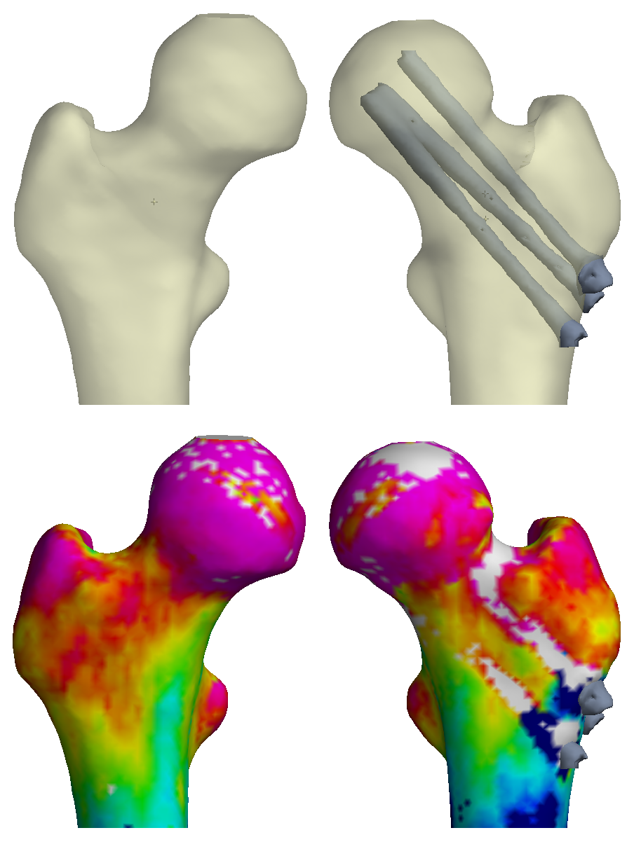
This is also relevant for patients with some sort of femur revision: here is one with some metal pins inserted. Again, can we use the un-revised side but then correct it where the metal has been inserted? |
| |
|
|
Hip fracture is a major issue affecting millions of people annually. We have recently been involved in research which has the potential to contribute significantly to our understanding of why hips fracture and what can be done to prevent them from doing so. This has mainly focussed on assessing various properties of the denser layer of cortical bone surrounding the less dense bone in the centre. We would like to extend this analysis to fractured bones and those with metalwork inserted, but there are two problems: they are very difficult to segment, and we need to relate locations on the fractured/metalwork side to those on the other femur.
Can these problems both be solved by using the good femur to segment the bad? If we take the surface of the good femur, flip it (e.g. to change from left to right) then 'fracture' it and align the bits correctly, then we can compare parameters (such as the thickness of the cortex, or outer bone layer) between the two sides.
This project contains plenty of interesting issues relating to computer graphics and computational geometry. How do we interact with the femoral surface in order to 'fracture' it in the right places? How do we align the bits correctly? How should this all be visualised? It would be suitable for anyone interested in creating and implementing techniques in computer graphics and computational geometry.
Click here for other medical imaging projects offered by Graham Treece.