Main > GMT_4YP_17_2
Dr Graham Treece, Department of Engineering
F-GMT11-2: Cleaning up surface meshes derived from CT data
 |  |  |
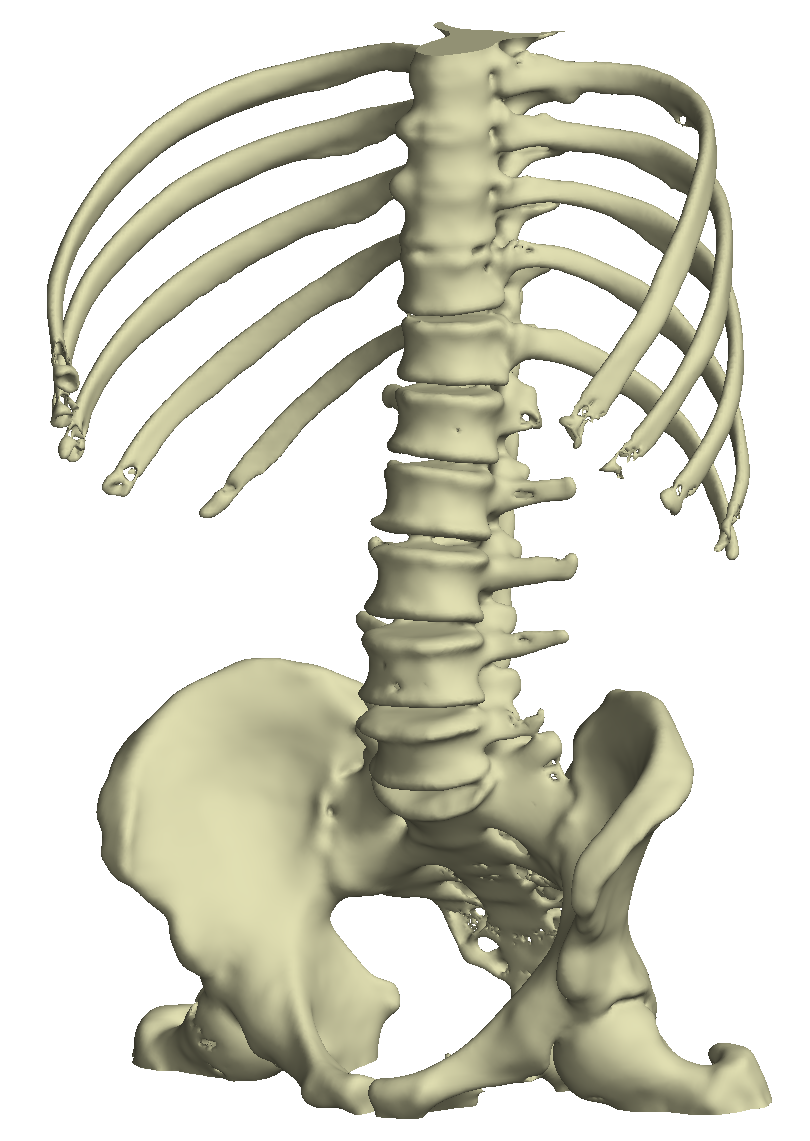
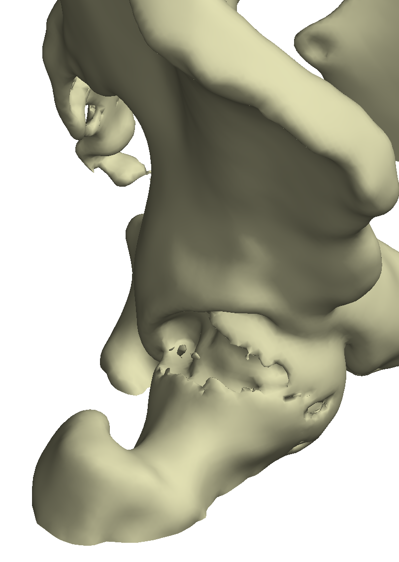
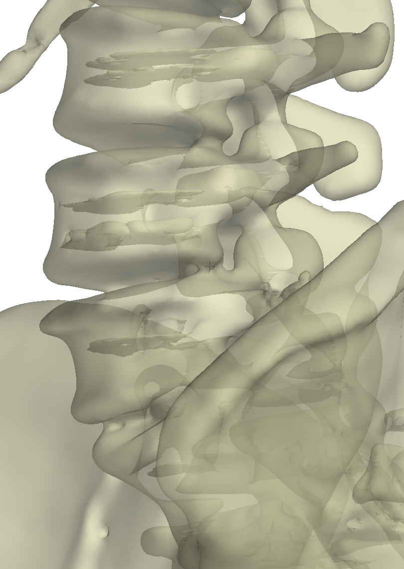
| This is a typical bone surface which has been automatically extracted by intelligently thresholding medical CT data. It is quite detailed, mostly correct, and useful for visualisation and as a framework for all sorts of subsequent measurements. | There are local errors though - here the top of the femur (femoral head) was very thin and a part of it was missed out during thresholding. Instead, the surface outlines some internal structures which we are not interested in. | In this transparent view the vertebrae have slight holes which have lead to internal 'planes' in the surface. These seem obviously wrong to our eyes: to what extent can these by cleaned up automatically by processing the mesh? |
Mesh extraction from medical data is a relatively common technique. Though it can be used for visualisation of surfaces within the data, it is particularly useful as a framework for subsequent measurement and analysis, for instance assessing bone cortical or mechanical properties. Such techniques need a good quality mesh which matches the specific data well, but they also need the mesh to have reasonable properties such as smoothness and topological consistency, as well as consistently representing the same (presumably outer) bone surface. Whilst there are techniques to improve mesh extraction in the first place, it is the aim of this project to see how much can be done to improve meshes once they have been created.
There are many algorithms out there for smoothing or 'correcting' surface meshes in some way, and quite a few have been implemented in a variety of free software applications. Hence the project will start with a substantial survey of existing techniques applied to this particular problem, to try to see what is possible, why it works, and what the current limitations are. The remainder of the project will try to fine tune or improve on these techniques for the specific task of removing features such as those shown in the figure above.
This is an algorithmic development / computational geometry / software project, so in the latter stages of the project experience of writing software will be essential, though the development could also be done using Matlab.
Click here for other medical imaging projects offered by Graham Treece.


