Main > GMT_4YP_20_2
Dr Graham Treece, Department of Engineering
F-GMT11-2: Mapping bone changes after osseointegrated implantation
 |  |  |
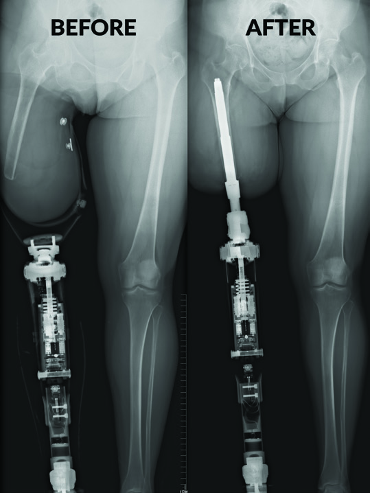
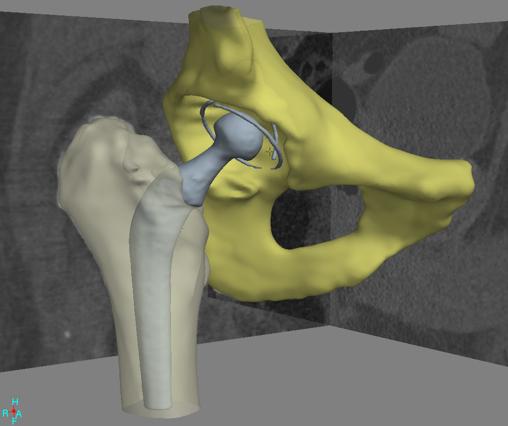
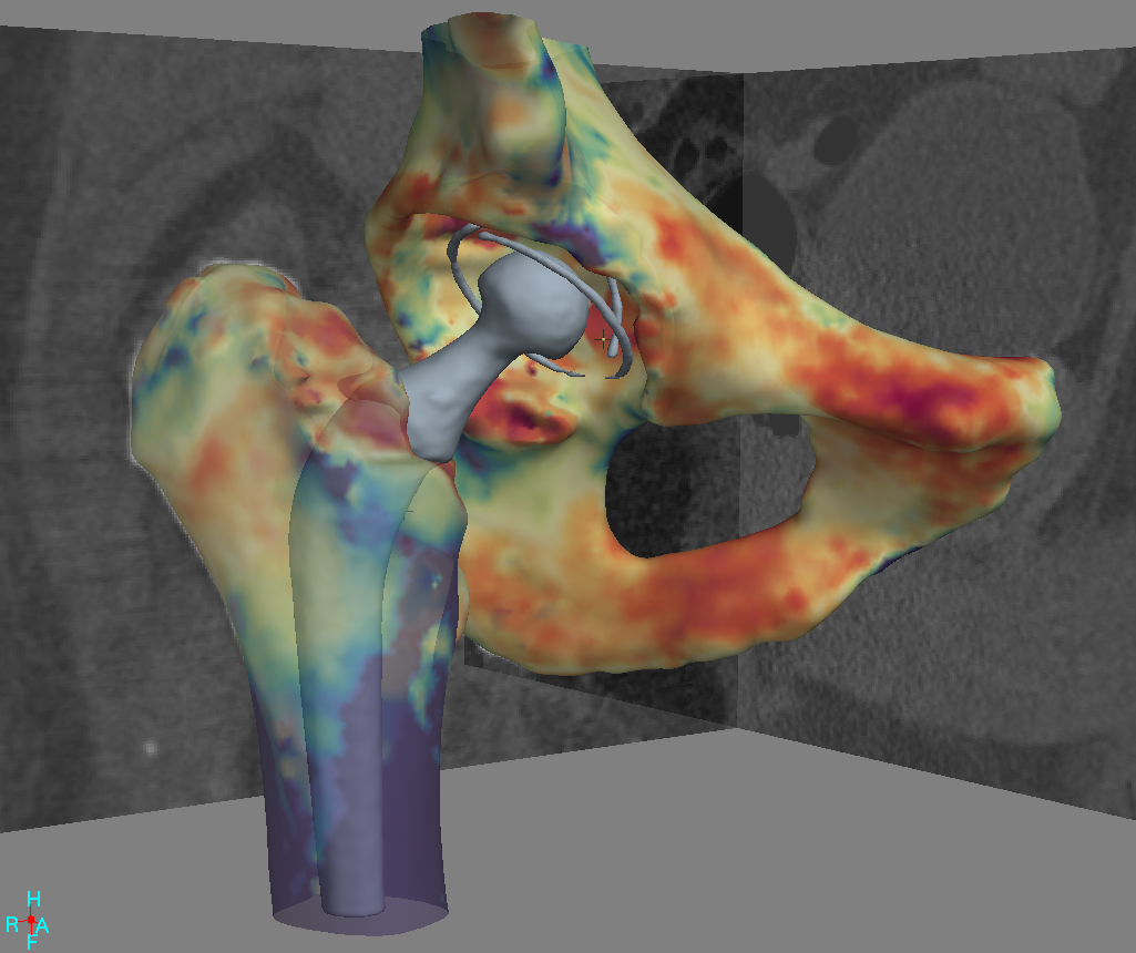
| Osseointegration is where the implant is directly attached to an existing bone (as on the right, above, image from Osseointegration Group of Australia) rather than being attached by a cup which is worn over the remaining leg (left, above) | It is possible to reconstruct both bone and metal surfaces using Computed Tomography X-ray (CT) data, here shown for a hip implant | Cortical Bone Mapping (CBM) is a technique to precisely measure the bone: the colour shows cortical thickness. Can this technique be used to investigate how the bone adapts to such implants? |
Osseointegrated implants are a relatively new technique by which a limb replacement is directly attached to an existing bone, rather than held on via a cup over the residual soft tissue. These have many potential benefits, and it is known that indeed people reliably demonstrate osseointegration of the bone into such an implant. That means the bone is biologically responding and growing into the implant. However, it would be good to have better knowledge of 1) how does the cortical bone respond (increased density, increased thickness, localized remodeling, does the bone increase these factors only at specific point loading areas or does the load distribute well along the length of the implant and bone?) in response to the implant, 2) over what time do these changes tend to occur, and 3) are there at-risk areas due to implant positioning or geometry?
This has currently been assessed using DEXA, a 2D technique which is essentially a carefully calibrated X-ray. It is possible to get a much fuller picture of the bone from Computed Tomography (CT, effectively a 3D X-ray). In particular, Cortical Bone Mapping (CBM) is a recent development which allows very precise assessment of bone from CT data. In CBM, thousands of measurements are made over the cortex (outer surface) of the bone, of cortical thickness, density, mass, and the underlying bone density. These measurements are very accurate and can be compared over time to show relatively minor localised changes in the bone.
In this project, CBM will be investigated as a tool to answer some of the interesting questions regarding osseointegrated implants, by making use of pre- and post- implant CT data sets from patients who have undergone this procedure. The project involves collaboration with Jason Hoellwarth, an orthopaedic surgeon who has contacts with the Osseointegration Group of Australia (see
http://www.osseointegration.org/).
This is an algorithmic development / computational geometry / software project, so experience of writing software would be useful, though much of the development could be done using existing in-house software research tools.
Click here for other medical imaging projects offered by Graham Treece.


